50
HIC
Monoclonal antibodies
Hydrophobic interaction is a very useful technique for the
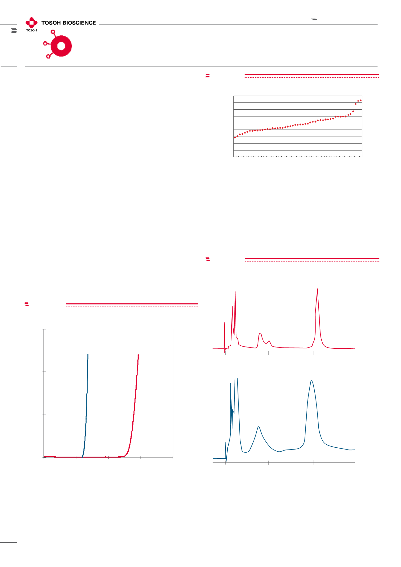
purification of monoclonal antibodies. The diverse hydro-
phobic nature of mAbs is seen in Figure 14. This figure
measures the hydrophobicity (using elution time as
a
surrogate measurement) of 51 different mouse IgGs on a
TSKgel Phenyl-5PW analytical column. Some of the IgGs
have elution times 2-3 times longer than others indicating
greater hydrophobicity. The TOYOPEARL series of HIC
ligands (Figure 2, page 33) with their different hydrophobic-
ities gives chromatographic developers a range of options
for finding the right ligand for their target molecule.
For a very hydrophobic mAb, such as mouse anti-chicken
14 kDa lectin, the less hydrophobic TOYOPEARL Ether
ligand works quite well. The purification from ascites fluid
(Figure 15) was performed with a 10 µm TSKgel Ether-5PW
semi-preparative column. Identical selectivity for scale-up
was found with corresponding 65 µm TOYOPEARL Ether-
650M resin.
hydrophobic interaction
chromatography
figure15
Purification of mAbs from ascites fluid
A. 10 µm TSKgel Ether-5PW
A. TSKgel Ether-5PW, 7.5mm ID x 7.5cm
B. Toyopearl Ether-650M, 7.5mm ID x 7.5cm
anti-chicken 14 kDa lectin,diluted ascites fluid,
A. 1.5mg in 100µL; B. 0.76mg in 50µL
60 min linear gradient from 1.5mol/L to 0mol/L (NH
4
)
2
SO
4
in 0.1mol/L phosphate buffer (pH 7.0)
136cm/h
UV @ 280nm
Column:
Sample:
Elution:
Linear velocity:
Detection:
Minutes
0
15
30
B. 65 µm Toyopearl Ether-650M
0
15
30
Minutes
mAb
mAb
Column: A. TSKgel Ether-5PW, 7.5 mm ID x 7.5 c L
B. TOYOPEARL Ether-650M, 7.5 mm ID x 7.5 cm L
Sample: anti-chicken 14 kDa lectin, diluted ascites fluid,
A. 1.5 mg in 100 µL; B. 0.76 mg in 50 µL
Mobile phase: 60 min linear gradient from 1.5 mol/L to 0 mol/L (NH
4
)
2
SO
4
in 0.1 mol/L phosphate buffer (pH 7.0)
Linear vel city: 136 cm/h; Detection: UV @ 280 nm
Purification of mAbs from ascites fluid
figure 13
Toyopearl Phenyl-600M Breakthrough curve (lysozyme)
Phenyl-600M
58
Phenyl-650M
27
Column:
7.8mm ID x 20cm
Sample:
1g/L lysozyme in 0.1mol/L
phosphate buffer (pH 7.0) + 1.8mol/L (NH
4
)
2
SO
4
Linear velocity: 300cm/hr
Detector:
UV @ 280nm
0
0.05
0.1
0.15
0
20
40
60
80
C/C 0
Lysozyme loaded (g/L)
Phenyl-650M
Phenyl-600M
Binding capacity (g/L)
(10% Breakthrough)
Binding capacity (g/L)
(10% Breakthrough)
Phenyl-600M
58
Phenyl-650M
27
Column: 7.8 mm ID x 20 cm L; Sample: 1 g/L lysozyme in
0.1 mol/L phosphate buffer (pH 7.0) + 1.8 mol/L (NH
4
)
2
SO
4
Linear velocity: 300 cm/h; Detector: UV @ 280 nm
TOYOPEARL Phenyl-600M Breakthrough curve (lysozyme)
figure14
0
10
20
30
40
50
60
70
80
90
No.1
No.3
No.5
No.7
No.9
No.11
No.13
No.15
No.17
No.19
No.21
No.23
No.25
No.27
No.29
No.31
No.33
No.35
No.37
No.39
No.41
No.43
No.45
No.47
No.49
No.51
Antibody
Elution time of each antibody
on TSKgel phenyl-5PW(min)
Column: TSK-GEL Phenyl-5PW
Eluent : (A) 0.1mol/L phosphate buffer containing 1.8mol/L ammonium
sulfate (pH 7.0)
(B) 0.1mol/L phosphate buffer (pH 7.0)
Flow Rate : 1 mL/min
Gradient : (B) 0% (0min)--0% (5min)--100% (65min) linear
Detector: UV @ 280nm
Samples : 51 kinds of mouse monoclonal antibodies
Hydrophobic diversity of mouse monoclonals
Plot of chromatographic elution times for 51 different mouse mAbs
Plot of chromatographic elution times for 51 different mouse mAbs
Column: TSKgel Phenyl-5PW; Mobile phase: (A) 0.1 mol/L phos-
phate buffer containing 1.8 mol/L ammonium sulfate (pH 7.0);
(B) 0.1 mol/L phosphate buffer (pH 7.0);
Flow rate: 1 mL/min; Gradient: (B) 0 % (0 min)-0 % (5 min)-100 % (65 min)
linear; Detector: UV @ 280 nm; Sa ples: 51 kinds of mouse monoclonal
antibodies
Hydrophobic diversity of mouse monoclonals


