9
IEC
IEC
TSKgel STAT SERIES
Applications with TSKgel STAT Cation Exchange
Columns
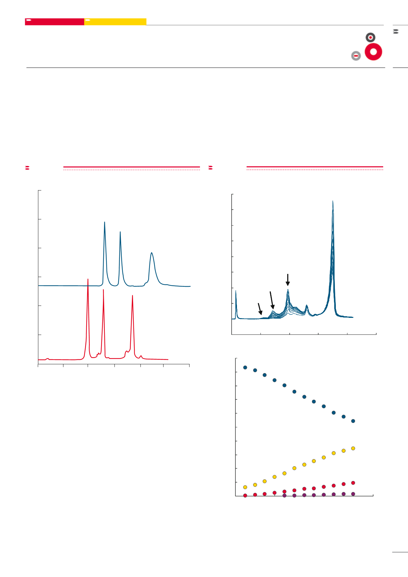
Fast Separations
The fast separation of protein standards was investigated
usingshort cationexchange columns (seeFigure7). ATSKgel
SP-STAT column shows superior resolution, better peak
shape, and shorter analysis time (< 60 seconds) compared
to a competitive monolithic SP-type column.
Reaction Monitoring
A sample of
β
-lactoglobulin (5 mg/mL) was reacted with
polyethylene glycol (5 kDa) in a pH 6.5 phosphate buf-
fer. The formation of PEGylated protein reaction prod-
ucts was monitored in 5 minute intervals on a 3.5 cm L
TSKgel SP-STAT column. As demonstrated in Figure 8,
peak areas of mono-, di-, and tri- PEGylated
β
-lactoglobulin
increased with reaction time, while the area of unreacted
β
-lactoglobulin declined.
(A)
(B)
1
2
3
Retention time (min)
0
0.2
0.4
0.6
0.8
1
1.2
mV
Columns:
A: TSKgel Q-STAT, 10µm, 3.0mm ID x 3.5cm
B: ProSwift SCX-1S Monolith, 4.6mm ID x 5cm
Eluent:
A: 20mmol/L sodium acetate (pH5.0)
B: 1.0mol/L NaCl in buffer A (pH5.0) for column A
1.5mol/L NaCl in buffer A (5.0) for column B
Gradient:
0% B (0min), 100% B (1min)
Flow rate:
A: 2.0mL/min
B: 4.73mL/min
Detection:
UV@280nm
Samples:
1. alpha-chymotrypsinogen A
2. cytochrome C
3. lysozyme
figure 7
Column: A: TSKgel SP-STAT, 10 µm, 3.0 mm ID x 3.5 cm L;
B: Competitor column 4.6 mm ID x 5.0 cm L
Eluent: A: 20 mmol/L sodium acetate (pH 5.0); B: 1.0 mol/L NaCl in buffer
A (pH 5.0) for column A; 1.5 mol/l NaCl in buffer A (pH 5.0) for column B;
Gradient: 0% B (0 min), 100% B (1 min);
Flow rate: A: 2.0 mL/min; B: 4.73 mL/min; Detection: UV @ 280 nm;
Samples: 1.
α
-chymotrypsinogen A; 2. cytochrome C; 3. lysozyme
5
10
15
20
25
30
35
40
45
50
0
0.5
1
1.5
2
2.5
UV @ 280 nm (1 Abs/1000 mV)
Mono- PEG
Di-PEG
Tri -PEG
Native
Retention time (min)
0
10
20
30
40
50
60
70
80
90
100
0
10
20
30
40
50
60
70
Reaction time (min)
Column:
A: TSKgel SP-STAT, 10µm, 3mm ID x 3.5cm
Eluent:
A: 20mmol/L sodium acetate buffer (pH5.0)
B: 1.0mol/L NaCl in buffer A (pH5.0)
Gradient:
0% B (0min), 100% B (2min)
Flowrate:
2.0mL/min
Detection:
UV@280nm
Sample:
pegylated
β
-lactoglobulin
Peak %
Native
Di -PEG
Tri- PEG
Mono-PEG
figure 8
Column: TSKgel SP-ST T, 10 µm, 3.0 mm ID x 3.5 c L; Eluent: A: 20 mol/L
sodi m acetate (pH 5.0); B: 1.0 mol/L NaCl in buffer A (pH 5.0);
Gradient: 0% B (0 min), 100% B (2 min); Flow rat : 2.0 mL/min;
Detection:UV @ 280 nm; Samples: PEGylated
β
-lactoglobulin
Fast protein separation
Pegylation monitoring


